The incredible images below show a single strand of DNA being pulled apart and read by the molecular machinery within a cell. The series, published in Nature as part of a study investigating how this machinery works, will be used to design new cancer-fighting drugs.
In order to snap the astonishing pictures with such fine detail, the researchers used a novel form of electron microscopy called Cryo-EM. The technique involved freezing the samples to -180°C (-292°F) to enable a resolution down to individual atoms. In fact, so impressive is this new method that pioneers of this technique were awarded the 2017 Nobel Prize for Chemistry.
“We used [the] really exciting new type of microscopy called Cryo-EM to do something no scientists have been able to do before,” explains Dr Alessandro Vannini, who led the research carried out at The Institute of Cancer Research, in a statement. “We were able to freeze and catch the RNA polymerase III complex in the act of attaching to, separating and reading the DNA code.”
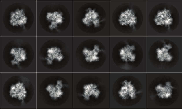
(They might not look like much to many of us, but quite amazingly these show a DNA strand being transcribed)
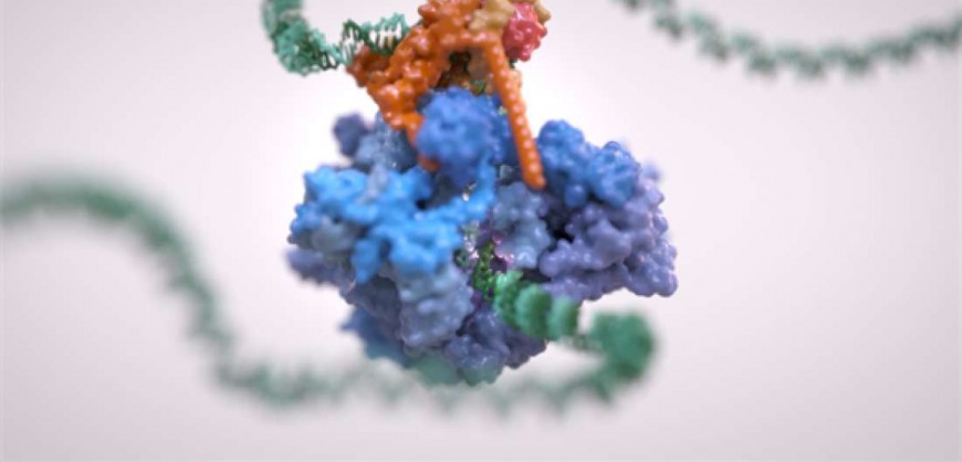
The protein complex RNA polymerase III is present in almost every cell in your body – as well as that of all other animals and plants – and is crucial for life to exist. It takes a strand of DNA, unzips it, and then reads the nucleotide bases to produce the building blocks necessary for the cell to grow and divide.
To better understand how components of the complex and accessory molecules interact and communicate with each other, the team managed to capture it the moment it was binding with a strand of DNA. The Cryo-EM enabled the researchers to see to a resolution roughly one-20,000th of the width of a human hair – and the image is stunning.
When cells become cancerous, they break free of the usual constraints that prevent cells from continually replicating themselves, including the complexes that copy the DNA. It is therefore hoped that by understanding exactly how the RNA polymerase III complex works, it can shed some light on how tumors grow.
“This beautiful study has unveiled a fundamental cog in the inner workings of cells, and one that is often exploited by cancers,” says Professor Paul Workman, the chief executive of the ICR. “It’s a hugely important finding in cell biology, and I hope that in future it will lead to new treatments for cancer patients.”
The imaging could help the researchers design drugs that turn the system on or off, or even break them apart completely, and so potentially provide a new way to stifle the growth of tumors.
Source: iflscience.com