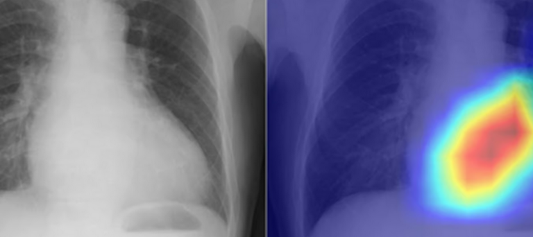
Researchers have used a deep-learning AI model to turn the humble chest X-ray into a more powerful tool for diagnosing heart problems. They say their novel approach could be used as a quick and accurate way of assessing heart function and checking for disease.
Chest X-rays are the most frequently conducted radiological test in the world and a common way for health professionals to diagnose lung and heart conditions. But, while they’re quick and easy to perform, an X-ray is a static image that’s unable to provide information about how the heart is functioning. For that, you need an echocardiogram.
Russian hackers lured diplomats in Ukraine with cheap BMW ad
An echocardiogram – commonly called an ‘echo’ – assesses how effectively the heart is pumping and whether the valves between the heart chambers are leaky or diseased. If the heart valves are diseased, the heart can’t pump blood effectively and has to work harder, which can lead to heart failure or sudden cardiac arrest and death. However, echocardiography requires a technician with specialized skills.
Read more: New Atlas
Ask me anything
Explore related questions