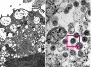
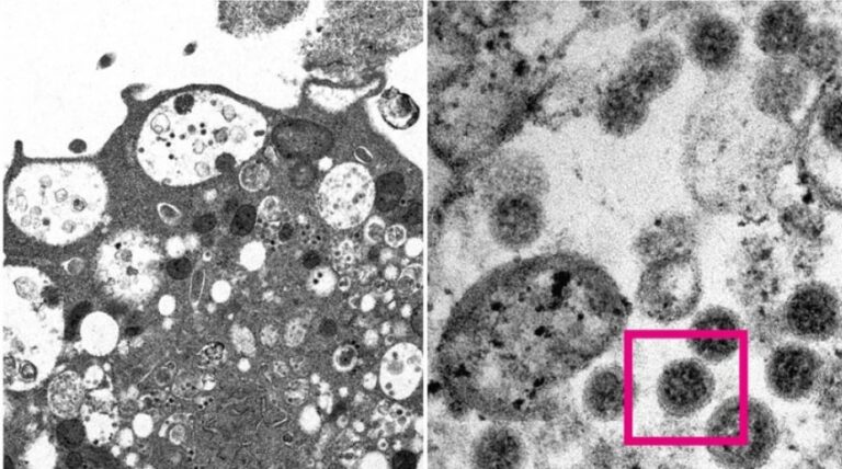
In a major discovery, scientists have unveiled the first microscopic image of the Omicron variant of the novel coronavirus that has triggered fears of another COVID-19 wave in the world. While the World Health Organisation (WHO) designated B.1.1.529 or Omicron, as a ‘variant of concern’, the University of Hong Kong on Wednesday released the image obtained by using an electron microscope.
The team of medical scientists including pathologists and virologists were able to take an electron micrograph of a monkey kidney cell (Vero E6) after it was infected with the Omicron variant of SARS-CoV-2. Both high and low-resolution Omicron images were released.

At low magnification, the image shows that the damage was done to cells with swollen vesicles containing small black viral particles, the researchers explained. Additionally, the high magnification revealed aggregates of the viral particles with corona-shaped spikes on the surface.
READ MORE
GSK Vir drug works against all Omicron mutations, new data shows
Sputnik has stated that the researchers at the Department of Microbiology at the University of Hong Kong managed to isolate Omicron from clinical samples, which will allow the development and production of vaccines against the new strain which has more mutations than any other known variant.
source republicworld.com
also read
The International Space Station (ISS) to be visible over parts of Greece
Ask me anything
Explore related questions


